Dinophysis acuminata Claparède & Lachmann
Classification:
Empire Eukaryota
Kingdom Protozoa
Subkingdom Biciliata
Infrakingdom Alveolata
Phylum Myzozoa
Class Dinophyceae
Order Dinophysiales
Family Dinophysiaceae
Genus Dinophysis
Pictures:
click on thumbnail for larger version.

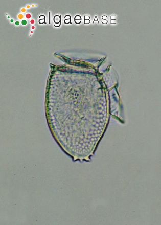
The characteristics of this genus such as the high girdle with lists giving the form of a collar, the flattened cell with the sulcus running down the right hand edge of the picture, also with a list giving the form of a sail, make it relatively easy to id. © Robin Raine.

Poulsbo, Washington, USA; Liberty Bay Marina, 400x, DIC. 30 May 2009. Karl Bruun. © Karl Bruun.
Dinophysis acuminata Claparède & Lachmann Poulsbo, Washington, USA; Liberty Bay Marina, 400x, DIC
Publication details
Dinophysis acuminata Claparède & Lachmann 1859: 408, pl. 20: fig. 17
Original publication: Claparède, É. & Lachmann, J. (1859). Études sur les infusoires et les rhizopodes. Mémoires de l'Institut National Genevois 6: 261-482.
Download PDF
Type species
The type species (holotype) of the genus Dinophysis is Dinophysis acuta Ehrenberg.
Status of name
This name is of an entity that is currently accepted taxonomically.
Origin of species name
Participle A (Latin), acuminate, i.e. tapering gradually or abruptly from inwardly curved sides into a narrow point (Stearn 1973).
Heterotypic Synonym(s)
Dinophysis ellipsoides Kofoid 1907
Dinophysis lachmannii Paulsen 1949
Dinophysis skagii Paulsen 1949
Dinophysis borealis Paulsen 1949
Dinophysis boehmi Paulsen 1949
Dinophysis lachmanii Solum 1962
General environment
This is a Marine species.
Usage notes
Faust & Gulledge (2002:24) notes this species has been found to produce okadaic acid causing diarrhetic shellfish poisoning (DSP). Please consult original reference for further details.
Harmful species
Type information
Type locality: North Sea: Norway (Faust & Gulledge 2002: 24). Holotype: (Faust & Gulledge 2002: 24).
Detailed distribution with sources
(as Dinophysis acuminata Claparède & Lachmann)
Europe: Britain (Parke & Dixon 1976), Croatia (Vilicic et al. 2009), Norway (Edvardsen et al., 2003), Romania (Caraus 2002), Sweden (Rehnstam-Holm, Godhe & Anderson 2002, Janson & Granéli 2003).
Atlantic Islands: Canary Islands (Gil-Rodríguez et al. 2003).
North America: Nova Scotia (Kim, Garbary & McLachlan 2004).
(as Dinophysis lachmannii Paulsen)
Europe: Britain (Parke & Dixon 1976).
(as Dinophysis skagii Paulsen)
Europe: Britain (Parke & Dixon 1976).
Key references
Dodge, J.D. (1982). Marine Dinoflagellates of the British Isles. pp. 303. London: Her Majesty's Stationery Office.
Edvardsen, B., Shalchian-Tabrizi, K., Jakobsen, K.S., Medlin, L.K., Dahl, E., Brubak & Paasche, E. (2003). Genetic variability and molecular phylogeny of Dinophysis species (Dinophyceae) from Norwegian waters inferred from single cell analyses of rDNA. Journal of Phycology 39: 395-408.
Faust, M.A. & Gulledge, R.A. (2002). Identifying harmful marine dinoflagellates. Contributions from the United States National Herbarium 42: 1-144.
Hallegraeff, G.M., Anderson, D.M. & Cembella, A.D., Eds (2003). Manual on harmful marine microalgae. Paris: UNESCO.
Janson, S. & Granéli, E. (2003). Genetic analysis of the psbA gene from single cells indicates a cryptomonad origin of the plastid in Dinophysis (Dinophyceae). Phycologia 42: 473-477.
Kim, K.Y., Garbary D.J. & McLachlan, J.L. (2004). Phytoplankton dynamics in Pomquet Harbour, Nova Scotia: a lagoon in the southern Gulf of St. Lawrence. Phycologia 43: 311-328.
Parke, M. & Dixon, P.S. (1976). Check-list of British marine algae - third revision. Journal of the Marine Biological Association of the United Kingdom 56: 527-594.
Steidinger, K.A. & Tangen, K. (1996). Dinoflagellates. In: Identifying Marine Phytoplankton. (Tomas, C.R. Eds), pp. 387-584. San Diego: Academic Press.
NCBI Nucleotide Sequences
Created: 07 May 2002 by M.D. Guiry
Verified by: 01 July 2010 by Wendy Guiry
Accesses: This record has been accessed by users 1201 times since it was created.
References
(Please note: only references with the binomials in the title are included. The information is from the Literature database.)
Hewes, C.D., Mitchell, B.G., Moisan, T.A., Vernet, M. & Reid, F.M.H. (1998). The phycobilin signatures of chloroplasts from three dinoflagellate species: a microanalytical study of Dinophysis caudata, D. fortii and D. acuminata (Dinophysiales, Dinophyceae). Journal of Phycology 34: 945-951, 3 figs.
Imai, I. & Nishitani, G. (2001 '2000'). Attachment of picophytoplankton to the cell surface of the toxic dinoflagellates Dinophysis acuminata and D. fortii (Research Note). Phycologia 39: 456-459.
Jacobson, D.M. & Andersen, R.A. (1994). The discovery of mixotrophy in photosynthetic species of Dinophysis (Dinophyceae): light and electron microscopical observations of food vacuoles in Dinophysis acuminata, D. norvegica and two heterotrophic dinophysoid dinoflagellates. Phycologia 33: 97-110, 52 figs.
Velo-Suárez, L. & Gutierrez-Estrada, J.C. (2007). Artificial neural network approaches to one-step weekly prediction of Dinophysis acuminata blooms in Huelva (Western Andalucía, Spain). Harmful Algae 6: 361-371.
Velo-Suárez, L., González-Gil, S., Gentien, P., Lunven, M., Bechemin, C., Fernand, L., Raine, R. & Reguera, B. (2008). Thin layers of Pseudo-nitzschia spp. and the fate of Dinophysis acuminata upwelling-downwelling cycle in a Galician Ría. Limnol. Oceanogr. 53(5): 1816-1834.
Vesk, M., Dibbayawan, T.P. & Vesk, P.A. (1996). Reserch Note : Immunogold localization of phycoerythrin in chloroplasts of Dinophysis acuminata and D. fortii (Dinophysiales, Dinophyta). Phycologia 35: 234-238, 8 figs.
Zingone, A., Montresor, M. & Marino, D. (1998). Morphological variability of the potentially toxic dinoflagellate Dinophysis sacculus (Dinophyceae) and its taxonomic relationships with D. pavillardii and D. acuminata. European Journal of Phycology 33: 259-273, 62 figs, 3 tables.


