Champia farlowii M.K.Griffith, C.W.Schneider & C.E.Lane 2017
Current name:
Champia farlowii M.K.Griffith, C.W.Schneider & C.E.Lane
Rhode Island, Charleston, Quonochontaug Central Beach; holotype - 01 May 2017. C.W. Schneider
Publication Details
Champia farlowii M.K.Griffith, C.W.Schneider & C.E.Lane 2017: 83, figs 3-10 [holotype fig. 4]
Published in: Griffith, M.K., Schneider, C.W., Wolf, D.I., Saunders, G.W. & Lane, C.E. (2017). Genetic barcoding resolves the historically known red alga Champia parvula from southern New England, USA, as C. farlowii sp. nov. (Champiaceae, Rhodymeniales). Phytotaxa 302(1): 77-89, 10 figs.
Type Species
The type species (holotype) of the genus Champia is Champia lumbricalis (Linnaeus) Desvaux.
Status of Name
This name is of an entity that is currently accepted taxonomically.
Type Information
Type locality: "Northwestern Atlantic Ocean, United States, Rhode Island, Charleston, Quonochontaug Central Beach, 41°20'12.8" N, 71°42'06.8" W"; (Griffith & al. 2017: 84) Holotype: C.W. Schneider (CWS) 15-27-2; 21 July 2015; shallow subtidal and drift; MICH; EC059 Notes: Isotype: EC060, Herb. CWS.
General Environment
This is a marine species.
Description
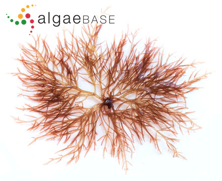
Plants reddish-brown to pink and greenish-red, erect to 5–8 cm tall, upright axes arising from small discoidal holdfasts; axial segments terete, barrel-shaped and constricted at septal regions, 1.0–1.5 diameters long in mature portions decreasing in length and width distally; branching mostly at septa, occasionally from internodal regions, mostly alternate but often opposite or irregular, frequently with multiple branches arising from the same node; main axes 1-2 mm in diameter, occasionally anastomosing with other branches; in surface view, septal cells in a single layer, angular and isodiametric, 36–95 µm long and 38–65 µm in diameter; cortex a single layer with two different cell sizes, larger cells irregularly polygonal to ovoid, 31–84 (–115) µm long and 20–42 µm in diameter, darkly staining smaller cells, subspherical to spherical, 10–22 (–27) µm in diameter, interspersed among them; thin longitudinal filaments 4–12 µm in diameter arising from where the septum join the cortical layer, running down the inner surface of the cortex surrounding the central mucilage-filled cavity of each segment, producing gland cells on the cavity side of the filaments, 5–6 µm in diameter; hair cells observed in some specimens originating from the small outer cortical cells; tetrahedral sporangia spherical 52–80 µm in diameter, scattered in the cortex, developing in mature segments several segments from the branch apices, abundant and obvious to the naked eye; mature cystocarps 500–845 µm in diameter and 650–910 µm tall, broadly ovoid to spherical and ostiolate at maturity, produced singly or in groups of 2–5 per node, tela arachnoidea present between the carposporophyte and pericarp wall; carpospores obpyriform, 23–40 µm in diameter and 57–75 µm long; spermatangia unknown.
Created: 01 May 2017 by M.D. Guiry.
Last updated: 30 January 2018
Verification of Data
Users are responsible for verifying the accuracy of information before use, as noted on the website Content page.
Linking to this page: https://www.algaebase.org/search/species/detail/?species_id=164214
Citing AlgaeBase
Cite this record as:
Craig Schneider in Guiry, M.D. & Guiry, G.M. 30 January 2018. AlgaeBase. World-wide electronic publication, National University of Ireland, Galway. https://www.algaebase.org; searched on 19 April 2024


